Highlights
Tomography. Fluorescence. Cryo.
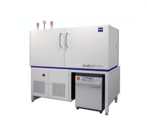
Xradia 800 Synchrotron: Hard X-ray nanotomography
3D tomographic imaging with X-rays provides detailed volumetric data of internal structures without the need for cutting or sectioning at the region of interest. Operating in the 5-11 keV energy range, Xradia 800 Synchrotron images a wide range of samples including battery and fuel cell electrodes, catalysts, and soft and hard tissue with resolution <30 nm. Xradia 800 Synchrotron is ideally suited for advanced techniques such as XANES spectro-microscopy for 3D chemical mapping and in situ imaging to enable you to study materials under real operating conditions.
Xradia 825 Synchrotron: Soft X-ray nanotomography
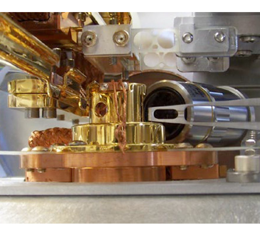
3D tomographic imaging in the soft X-ray range, including the water window up to about 2.5 keV, is ideally suited for structural imaging of whole cells and tissue. Cryogenic sample handling enables you to image in a frozen hydrated state, minimizing effects of radiation damage while maintaining the sample as close to its natural state as possible. Further applications include chemical state mapping of both organic and inorganic materials and imaging of magnetic domains.
Benefits
With ZEISS proven synchrotron platforms, invest your time and energy into your research rather than costly and time-consuming in-house development.
Choose the system that best suits your research needs on proven 3D X-ray microscopy platforms.
Xradia 800 Synchrotron: Hard X-ray nanotomography
- Leverage non-destructive 3D tomographic imaging<30 nm resolution for detailed volumetric data without the need for cutting or sectioning at the region of interest
- Experience flexibility for advanced techniques and image a wide range of samples in situ to understand the effect of real operating conditions with unparalleled image quality and throughput
- Map oxidation state in electrochemical devices or catalysts using XANES spectroscopy
Xradia 825 Synchrotron: Soft X-ray nanotomography
- Use the “water window” energy range to image organic materials in their natural, wet environment with high contrast
- Image the structure of whole cells and tissue, using the cryogenic sample handling to minimize the effects of radiation damage
- Correlate to optical fluorescence microscopy for combined imaging of structure and function
Applications
ZEISS Xradia Synchroton Family
| Xradia 800 Synchrotron | Xradia 825 Synchrotron | |
| Materials Science | Monitor battery electrode particles in operando during the charge-discharge cycle. Perform chemical imaging of catalyst particles in situ. Analyze SOFC nanostructure in situ at operating temperature. | Perform chemical imaging of polymers by spectro-microscopy. |
Life Sciences | Study toxicity of nanoparticles in cells and tissue. Image and quantify the nanostructure of bone. | Visualize ultrastructure in whole, unsectioned cells in the frozen hydrated state. Correlate X-ray and optical fluorescence microscopy for combined structural and functional imaging. |
Natural Resources, Geo- and Environmental Sciences | Visualize morphology of iron melt at Earth’s lower mantle conditions. Study microstructure of soil particles relevant to water retention. | Study micro-organisms in wet environments. |
Electronics | Image integrated circuits to find malicious modifications. | Image magnetic domains on the nanoscale. |





Đánh giá
Chưa có đánh giá nào.