


Highlights
Extending the Limits of Micro- and Nano-CT Solutions
- Non-destructive sub-micron scale microscopy of intact samples
- Higher flux and faster scans without compromising resolution
- True spatial resolution of 500 nm with a minimum achievable voxel size of 40 nm
- High resolution across a broad range of sample types, sizes, and working distances
- In situ imaging for non-destructive characterization of microstructures in controlled environments and over time
- Upgradeable and extendible with future innovations and developments
Highest Resolution and Flux
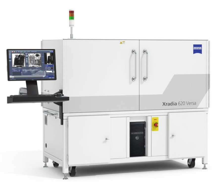
Where traditional tomography relies on single-stage geometric magnification, Xradia Versa features a combination of unique two-stage magnification optics and a high flux X-ray source to produce faster sub-micron scale resolution images. The Resolution at a Distance (RaaD) architecture enables high resolution 3D imaging of larger, denser objects including intact components and devices. The optional flat panel extension (FPX) enables rapid scans of very large samples (up to 25 kg), allowing navigation to interior regions of interest.
New Degrees of Freedom
Use the industry’s most comprehensive 3D X-ray imaging solution for advanced scientific and industrial research: Maximize absorption and phase contrast to achieve unprecedented characterisation of your materials and their properties. Unlock 3D crystallographic information with Diffraction Contrast Tomography. Improve scan speed and accuracy of large or irregular samples with advanced acquisition techniques. Apply machine learning algorithms to help with post-processing and segmentation of your samples.
Premier 4D / In Situ Solution
ZEISS Xradia 600-series Versa can non-destructively characterize the 3D microstructure of materials under controlled perturbations (in situ), and observe the evolution of structures over time (4D). By leveraging Resolution at a Distance, the Xradia Versa maintains highest resolution across large working distances, accommodating both sample, environmental chamber, and high precision in-situ load rigs without sacrificing resolution. The Versa seamlessly integrates with other ZEISS microscopes to solve multi-scale correlative imaging challenges.
Lithium Ion Batteries
Typical tasks and applications
- Recipe development and supply chain control: Inspection of intact samples for effective supplier control, revealing changes in recipe or cost savings that may affect performance or longevity
- Safety and quality inspection: Identification of debris, particle formation, burrs at the electrical contact or damage to the polymer separator
- Lifetime and aging effect: Longitudinal studies of aging effects
")
")
")

Electronics and Semiconductor Packaging
Typical tasks and applications
- Perform structural and failure analysis for process development, yield improvement and construction analysis of advanced semiconductor packages, including 2.5/3D and fan-out packages
- Analyze printed circuit boards for reverse engineering and hardware security
- Non-destructively image across length scales from module to package to interconnect for submicron-resolution characterization of defects at speeds that can complement physical cross-sectioning
- Enable better understanding of defect locations and distributions by viewing unlimited virtual cross-section and plan-view images from all desired angles


Additive Manufacturing
Typical tasks and applications
- Detailed shape, size, and volume distribution analysis of particles in Additive Manufacturing (AM) powder bed to determine proper process parameters
- High-resolution, non-destructive imaging for microstructural analysis of AM parts
- 3D imaging for comparison with the nominal CAD representation
- Detection of unmelted particles, high-Z inclusions, and voids
- Surface roughness analysis of inner structures that cannot be accessed by other methods
.")


Materials Research
Typical tasks and applications
- Characterize three-dimensional structure
- Observe failure mechanisms, degradation phenomena, and internal defects
- Investigate properties at multiple length scales
- Quantify microstructural evolution
- Perform in situ and 4D (time dependent studies) to understand the impact of heating, cooling, desiccation, wetting, tension, compression, imbibition, drainage and other simulated environmental studies


Raw Materials
Typical tasks and applications
- Perform multiscale pore structural and fluid flow analysis
- Directly measure fluid flow at the pore scale using in situ flow equipment
- Analyze crystal structures using LabDCT
- Particle analysis with full 3D reconstruction
- Advance mining processes: analyze tailings to maximize mining efforts; conduct thermodynamic leaching studies; perform QA/QC analysis of mining products such as iron ore pellets
- Understand grain orientations in steel and other metals


Life Sciences
Typical tasks and applications
- 3D imaging of biological samples in their natural surroundings
- Imaging of plant roots still embedded in their original soil without any special sample preparation
- Imaging of fragile animals and plants without any sample preparation and sectioning
- Sub-micron imaging of solid structures like seeds as a whole


Highest Resolution without Compromise
Standard X-ray computed tomography (CT) is limited to small sample sizes when imaging at high resolution; due to the geometric nature of magnification. Maintaining high resolution for larger samples is impossible due to the longer working distances required. High resolution imaging in CT systems also requires low X-ray flux, reducing the throughput of the measurement. This limits the practical application of maximum resolution claimed by most CT manufacturers.
ZEISS Xradia 600-series Versa overcomes these trade-offs by integrating dual-stage magnification architecture with high flux X-ray source technology.
ZEISS specifies true spatial resolution, bringing a standard measure of microscope performance to 3D X-ray measurement. Spatial resolution refers to the minimum separation at which two features can be resolved by an imaging system. ZEISS Xradia 600-series Versa systems obtain true spatial resolution of 500 nm with a minimum achievable voxel size of 40 nm.
Higher X-ray Flux Source
Numerous Advantages
ZEISS Xradia 600-series Versa introduces a breakthrough high power (25 W) X-ray source technology that can provide significantly higher X-ray flux compared to its predecessors. The new source pushes the boundaries of performance with improved thermal management, increased flux and throughput while preserving resolution performance. A new source control system improves source responsiveness enabling faster scan setup leading to an easier and more engaging user experience.
What higher X-ray flux offers:
- Faster tomography scans
- More sample runs
- More regions of interest
- Higher contrast-to-noise ratio
- Stronger diffraction patterns
- Enabling long/multi-scan workflows
(in situ, DSCoVer, stitching, DCT)


ZEISS X-ray Microscopes
The Versatile Advantage of RaaD
ZEISS Xradia Versa uses a two-stage magnification architecture to enable sub-micron resolution imaging at large working distances (Resolution at a Distance) for a diverse set of sample sizes and types. Images are initially magnified via geometric projection as with conventional microCT, the projected image is cast onto a scintillator, converting X-rays to a visible light image which is then optically magnified by microscope optics before acquisition by a CCD detector.
With more X-ray photons available, the ZEISS Xradia 600-series Versa provides faster time to results for the widest range of sample sizes and types, without compromising resolution.
Conventional microCT Architecture

ZEISS XRM Two-stage Magnification Architecture

Accessories
Extend the Range of Possibility for Advanced Material Characterization in 3D
LabDCT
Bringing synchrotron capabilities to your lab experiments
LabDCT (Diffraction Contrast Tomography), available exclusively on Xradia 620 Versa, enables non-destructive mapping of orientation and microstructure in 3D. Direct visualization of 3D crystallographic grain orientation opens up a new dimension in the characterization of metal alloys and polycrystalline materials.
- Obtain comprehensive 3D microstructure analysis from large volumes down to local individual grain boundary analysis.
- Investigate microstructural evolution with 4D imaging experiments.
- Combine 3D crystallographic information with 3D microstructural features.
- Combine modalities to understand structure-property relationships.

Flat Panel Extension
Image even larger samples with high throughput

Optional Flat Panel Extension (FPX) delivers large-sample, high throughput scanning with ZEISS best-in-class image quality. FPX enhances imaging flexibility and creates workflow efficiencies with an all-in-one system for industrial and academic research.
Scout-and-Zoom is a unique capability of ZEISS X-ray microscopes that leverages FPX to perform exploratory “Scout” scans across a large field of view to identify interior regions of interest for higher resolution “Zoom” scans without complex sample preparation
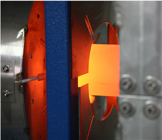
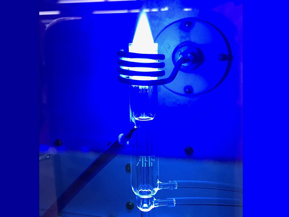
In Situ experiments
Push the limits for scientific advancement
ZEISS Xradia Versa provides the industry’s premier 3D imaging solution for the widest variety of in situ rigs, from high pressure flow cells to tension, compression and thermal stages.
To accommodate various types of in situ apparatus, such experiments require samples to be mounted further away from the X-ray source. With traditional microCT systems, this significantly limits the resolution achievable during such measurements. ZEISS X-ray microscopes are uniquely capable of Resolution at a Distance (RaaD) technology, which give the highest fidelity of 3D structural information during in situ imaging.

Autoloader
Increase your sample handling efficiency

Maximize your instrument’s utilization with the optional Autoloader, available for all instruments in the ZEISS Xradia Versa series. Reduce the frequency of user interaction and increase productivity by queueing multiple jobs. Load up to 14 sample stations, which can support up to 70 samples, and set to run overnight, or across multiple days. Unprecedented mechanical stability enables high volume quantitative repetitive scanning of like samples.
Wide Field Mode
Flexibly image larger samples

Wide Field Mode (WFM) can be used to image across an extended lateral field of view. The wide lateral field of view can provide 3x larger 3D volume for large samples, or give a higher voxel density for a standard field of view. All Xradia Versa systems are capable of WFM with the 0.4x objective. The Xradia 620 Versa system also features WFM with the 4x objective. In combination with Vertical Stitching, WFM enables you to image larger samples at exceptional resolution.
Automated Filter Changer
Simplify exploration of challenging samples
 offers 12 standard filters with room for 12 custom filters.")
X-ray source attenuation filters are used to tune the X-ray energy spectrum illuminating the sample to optimise contrast, which depends on the specific material properties of the sample. Every ZEISS Xradia Versa comes standard with a set of 12 filters. ZEISS Xradia 610 Versa is equipped with a single filter slot allowing for manual filter change. ZEISS Xradia 620 Versa systems feature an Automated Filter Changer (AFC), which improves ease of use, allowing seamless change of filters for convenient investigation of unknown samples.
The Xradia 600-series Versa
| ZEISS Xradia 610 Versa | ZEISS Xradia 620 Versa | |
|---|---|---|
| Spatial resolutiona | 500 nm | 500 nm |
| Resolution at a Distance (RaaD™)a,b (at 50 mm working distance) | 1.0 μm | 1.0 μm |
| Minimum Achievable Voxelc (Voxel size at sample at maximum magnification) | 40 nm | 40 nm |
| Source Voltage Range | 30–160 kV | 30–160 kV |
| Source Maximum Power Output | 25 W | 25 W |
| Scout-and-Scan™ Control System | ✓ | ✓ |
| Scout-and-Zoom | ✓ | ✓ |
| Vertical Stitch | ✓ | ✓ |
| XRM Python API | ✓ | ✓ |
| Automated Filter Changer (AFC) | ✓ | |
| High Aspect Ratio Tomography (HART) | ✓ | |
| Dual Scan Contrast Visualizer (DSCoVer) | ✓ | |
| Wide Field Mode | 0.4x | 0.4x and 4x |
| ZEISS LabDCT for Diffraction Contrast Tomography | Optional | |
| ZEISS Autoloader | Optional | Optional |
| In Situ Interface Kit | Optional | Optional |
| ZEISS OptiRecon | Optional | Optional |
| ZEISS ZEN Intellesis | Optional | Optional |
| ORS Dragonfly Pro | Optional | Optional |
a Spatial resolution measured with ZEISS Xradia 2D resolution target, normal field mode, optional 40x objective.
b RaaD™ working distance defined as clearance around axis of rotation
c Voxel is a geometric term that contributes to but does not determine resolution, and is provided here only for comparison.
ZEISS specifies resolution via spatial resolution, the true overall measurement of instrument resolution
Protect Your Investment
ZEISS X-ray microscopes are designed to be upgradeable and extendible with future innovations and developments to protect our customer’s investment. This ensures the microscope capabilities evolve with the advancements in leading edge technology.
From ZEISS Xradia Context microCT, to ZEISS Xradia 510/520 Versa, and now with the addition of ZEISS Xradia 610/620 Versa, users can field-convert their systems to the latest X ray microscopes.


Software
Create Efficient Workflows by Using The Simple Control System
Easily scout a region of interest and specify scanning parameters within the Scout-and-Scan Control System. Take advantage of the easy-to-use system in your central lab where users may have a variety of experience levels.
Benefit from:
- Internal camera for sample viewing
- Recipe control (set, save, recall)
- Multiple energies
- Multiple samples with Autoloader option
- Micropositioning capability with a simple mouse click

Reviews
There are no reviews yet.